Brain Terminology

A small hole(s) is/are created in the skull to remove blood or take samples of a brain tumor.

The narrowing of the carotid artery by a plaque of fatty deposits (arthrosclerosis), which reduces the blood flow to the brain. 

A thin or weak artery in the brain, which can bulge out and fill with blood. Aneurysms can spontaneously rupture, causing life-threatening consequences.

An abnormal connection between the arteries and veins which can be seen in the brain and spine. AVMs can rupture and cause life-threatening consequences.

The fluid that is by the brain to support and nourish the brain and spinal cord.

An abnormal accumulation of cerebrospinal fluid (CSF) that builds up within the fluid containing cavities (ventricles) of the brain, which can increase the intracranial pressure.

A general term for surgery which requires the use of a which is typically microscope.

Surgical Procedures for the Brain

A craniotomy that is performed while the patient is awake to participate for an exam, in order to monitor and preserve function. We are one of the few centers in the United States to conduct this type of surgery.

A surgical procedure where a metal clip is placed at the base of the aneurysm to prevent blood flow into the aneurysm.  The aneurysm clips are MRI compatible.

A surgical procedure to open and remove the fatty deposits in the carotid artery to reduce the risk of stroke.

A surgical procedure that removes part of the bottom of the back of the skull to create more room for the brainstem and promote normal cerebrospinal fluid (CSF ) flow at the brain and spine junction.

Computer Assisted Brain Surgery represents a surgical concept and set of methods that uses computer technology for surgical planning and for helping to guide the surgical approach. In computer-assisted brain surgery, the treatment team uses imaging technologies, such as magnetic resonance imaging (MRI), intraoperative MRI, computerized tomography (CT), to create a 3-D model of a person’s brain. The computer system precisely guides the brain surgeon (neurosurgeon) to plan the safest way to the area(s) of your brain that requires treatment.

A surgical procedure where part of the skull is removed and not replaced for weeks to months to allow room for the brain as it swells. Swelling of the brain can occur in victims of traumatic brain injury and stroke.

A surgical procedure to repair a bone defect that was removed from a previous operation or injury. The bone can be from the stored original skull piece or a synthetic bone substitute.

A surgical procedure that removes part of the skull to expose and gain access to the brain.

The stimulation of the deep brain structures is used to treat Parkinson’s disease, dystonia and Essential Tremors. DBS is recommended for patients with these conditions who are not being fully controlled by standard medications. Inova Medical Group neurosurgeons use state-of-the-art stereotactic imaging systems to visualize, analyze and plan for the best surgical approach using real-time interactive 3-D image guidance.

We are one of the few centers in the United States to conduct this type of surgery.

The intracranial electrodes are placed through a burr hole to determine the seizure focus (where the seizure occurred) in a patient with medically refractory complex partial seizures; such cases are consideration for NeuroPace, Responsive Neurostimulation. 

A surgical procedure used to relieve symptoms, such as facial pain or facial twitching, that can occur when the nerve is compressed by an artery or vein.

NeuroPace is a Federal Drug Administration (FDA) approved system as an adjunctive treatment (another treatment used together with the primary treatment) for adults with medically refractory partial seizures occurring from one or two seizure targets. These targets are identified by the neurosurgeon following electrode placement. NeuroPace is placed under the skull by our neurosurgeon.

A surgical procedure in which a craniotomy is performed, or a burr hole is created, to gain access inside the skull to remove blood (hematoma).  

A small tube surgically placed into the ventricles to help drain cerebrospinal fluid and redirect it to drain into abdomen (peritoneum). When draining to the abdomen is not an option, a ventriculoatrial (heart) or ventriculopleural (lung) shunt is placed. This is typically the case when there is hydrocephalus. 

VNS is approved by the Federal Drug Administration (FDA) to reduce seizure frequency for patients with partial onset seizures that do not respond to medical treatment. This procedure applies electrodes around the vagus nerve in the neck. The electrodes are connected to an implanted programmable generator.

Surgical Procedures for the Spine

A surgical procedure of the cervical spine that removes degenerative or herniated discs to relieve the spinal cord or nerve root compression. Typically, a small plate with screws and a cage are used to achieve fixation and fusion. 

This procedure approaches the lumbar vertebrae from the front of the abdomen to correct the alignment of the lumbar spine.  A general surgeon assists to expose the lumbar vertebrae.

ALIF is performed to remove a lumbar disc (discectomy) and fuse the spine.

There are a variety of bone graft substitutes available or are being evaluated in various stages of clinical trials for use in spine fusion surgery. In general, these types of bone graft are made of a synthetic or manipulated type of a naturally-occurring product.

A surgery involving removal of a generated or herniated cervical disc and replacing it with an artificial disc. It is done when the space between your vertebrae has become too narrow and part of your vertebrae or your cervical disc is pressing on your spinal cord or spinal nerves, causing you pain, numbness, or weakness. Candidates for this surgical approach are patients who must avoid fusion of the cervical spine.  

A cervical foraminotomy is a surgery performed to remove bone compressing on the exiting nerve to relieve arm pain.

Cervical fusion is a surgical procedure in which two or more vertebrae of the neck are joined or fused together. Fusion surgeries typically require the use of bone graft to facilitate fusion and screws and plates or rods to stabilize the cervical spine.

A corpectomy is a surgical procedure used to remove a vertebral body and adjacent vertebral discs in order to decompress the spinal cord and stabilize the spine.


A discectomy is the surgical removal of a herniated disc in the spine. A herniated disc can cause compression of the nerve root and cause pain, numbness, or weakness in the arms or legs. 

XLIF is a minimally-invasive fusion surgery of the lumbar spine in which the surgeon accesses the intervertebral disc space and fuses the lumbar spine (low back) from the side (lateral), rather than from the front (anterior) or the back (posterior). 

Kyphoplasty is a spinal procedure in which a balloon is deployed in a vertebral body fracture in order to give height to the vertebral body. Bone cement is then injected to fractured vertebra.

The goal of kyphoplasty is to relieve back pain caused by vertebral compression fractures.

Laminectomy is surgery that creates space by removing the lamina — the back part of the vertebra that covers your spinal canal. Also known as decompression surgery, laminectomy enlarges your spinal canal to relieve pressure on the spinal cord or nerves.

This is a procedure in which the lamina are hinged laterally to provide more room for the spinal cord and secure them into a new position using small screws.


This is a minimally invasive procedure used to treat leg or back pain generally caused by degenerative disc disease. The procedure is performed through the patient's side, avoiding the major muscles of the back.


Lumbar fusion is a surgical procedure in which two or more vertebrae of the lower back are joined or fused together. Fusion surgeries typically require the use of bone graft to facilitate fusion, and screws and rods to stabilize the lumbar spine.

This procedure is designed to remove a small portion of the bone over the nerve root and/or disc material from under the nerve root to give the nerve root more space, relieve pressure, and provide a better healing environment.

This is a surgical procedure in which a portion of a herniated nucleus pulpolsus — the jelly like substance in the middle of the spinal disc — is removed by way of a surgical instrument or laser, while using an operating microscope for magnification.

Evidence-based medicine is the driver for patient care at Inova. When spine surgery is required the surgeon will review procedure options that may be available. One of them might be minimally invasive spine surgery.

Compared to open spine surgery, minimally invasive spine surgery, sometimes referred to as MIS or MISS, accesses the spine through smaller incisions. These can be from 2-4 inches long compared to longer incisions in open approach spine surgery. Through the smaller incisions the surgeon is still able to move muscle tissue and expose the affected spine region.

The overall benefit to performing surgery with smaller incisions is for minimally invasive surgical approaches to be faster, safer and require less recovery time. Comparing this to open surgical approaches, there is reduced trauma to the muscles and soft tissues.

Some potential benefits to minimally invasive spine surgery include:

  • Small incisions result in less scarring
  • Reduction in soft tissue damage
  • Less injury to the muscles
  • Less post-operative pain
  • Reduced blood loss
  • Faster recovery

A note to understand: not all spine surgeries can be done with minimally invasive spine surgery techniques.

New techniques for minimally invasive spine surgery continue to evolve. Example conditions treated using minimally invasive surgical techniques include:

  • Degenerative disc disease
  • Herniated disc
  • Lumbar spinal stenosis
  • Spinal deformities such as scoliosis
  • Spinal infections
  • Spinal instability including spondylolisthesis
  • Vertebral compression fractures
  • Spinal tumors

Inova has the latest technologies for minimally invasive spine surgery with augmented reality and minimally invasive spine surgery with robotic instrumentation.

Minimally invasive surgery is the least invasive and effective surgical technique for treating spinal disc herniation patients. Spine surgeons use the microscope to remove the herniated disc.

This procedure approaches the lumbar vertebrae from the side of the abdomen to correct the alignment of the lumbar spine. A general surgeon assists to expose the lumbar vertebrae.

OLIF is performed to remove a lumbar disc (discectomy) and fuse the spine.

This procedure is used to treat patients with severe spinal curvature of the spine causing debilitating pain in the back or legs. The surgery requires multiple levels of the spine to be fused to correct the deformity of the spine.

This procedure relieves pressure of the nerves in the spine. Spinal decompression can be addressed with a laminectomy, foraminotomy or a discectomy.

This surgical procedure to implant titanium, titanium-alloy, or bone grafts into the spine. Instrumentation provides a permanent solution to spinal instability.

This procedure may include a biopsy, partial removal, or gross total resection of the spinal tumor. If the spinal tumor has eroded away the support structures of the spine, a fusion surgery may be required.

This procedure approaches the lumbar vertebrae from the back of the spine. 

TLIF may be performed to treat degenerative lumbar spine with instability

A surgical procedure involving the removal of part or all of the vertebral body to decompress the spinal cord or nerve roots of the spine.

Advanced Surgical Equipment and Technologies

CyberKnife System is a radiation therapy device used to deliver radiosurgery for the treatment of tumors in the brain and spine. The neurosurgeons and radiation oncologist use state-of-the- art imaging to plan the delivery of the radiation. The fully robotic CyberKnife delivers radiation safely by tracking patient’s anatomy and movement to avoid exposing radiation to healthy tissues. 

This is a neurosurgical technique that places electrodes deep in the brain to control abnormal impulses of the brain in patients with Parkinson’s disease, dystonia, and essential tremors who have unsuccessfully tried to control their symptoms with medications.

Learn More About DBS at Inova

An electroencephalography (EEG) and electromyography (EMG) are used during surgery to monitor the functional integrity of the brain or spinal nerves. This provides the surgeon real-time feedback allowing them to make adjustments in the surgical approach and treatment in order to reduce the risk of injury to the nervous system.

Laser Interstitial Thermal Therapy (LITT) 

A state-of-the-art MRI-guided, minimally invasive, laser ablation system with real-time monitoring of the brain tissue. LITT allows for treatment of deep brain tumors, which can be dangerous with traditional surgery. Using advanced computer imaging techniques, the laser is guided through the small inserted catheter with real-time MRI, and delivers thermal energy directly on tumor. 

Laser Ablation

Laser ablation neurosurgery is the latest technology in treatment of tumors. Neurosurgeons use an MRI-guided, high-intensity laser probe designed especially for the treatment of deep inoperable brain tumors allowing them to probe through the brain and into the tumor. Once inside the tumor, the probe discharges highly focused thermal energy to coagulate and kill cancer cells, while leaving surrounding brain tissue undamaged. 

This technology is Federal Drug Administration (FDA)-approved for neurosurgical procedures. Use of this technology was pioneered and adopted by two of Inova's neurosurgeons, Dr. Mateo Ziu and Dr. Mahesh Shenai. 

Meet Mateo Ziu, MD

Meet Mahesh Shenai, MD

surgeon performing surgery with augmented reality
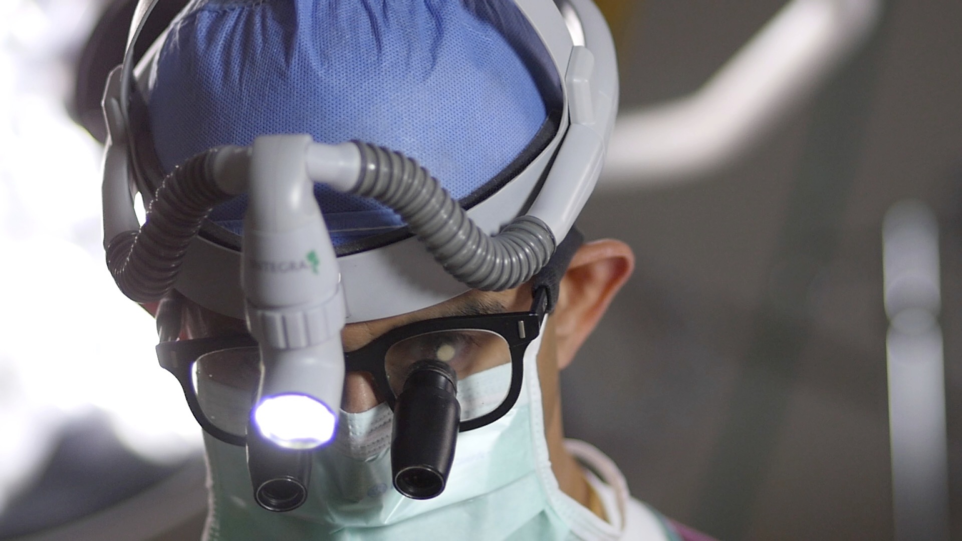
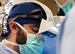
Augmented Reality (AR) guidance for spine surgery provides spine surgeons the ability to visualize the three-dimensional spine anatomy of a patient during surgery. This can be compared to working with "X-ray vision" glasses.

The benefit of this is to accurately navigate surgical instruments and implants while keeping focus directly on the patient, rather than a remote screen which had been the more common practice.

Augmented Reality spine surgery, like spine robots provides a safer, more efficient surgery. Having the aid of augmented reality guidance systems takes the best surgical navigation systems and improves upon them to meet the needs of spine surgeons and provide technical confidence in the operating room.

robotic assisted surgery
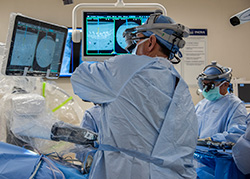
Robotic-assisted spine surgery is an advancement in spine surgery. It enables surgeons to plan their surgery even before their first patient cut. Once spine surgery begins, the robot provides screw placement with better accuracy due to the guidance system in the robot. Since the surgery was pre-programmed with prior imaging both the patient and operating room staff are subjected to less perioperative X-ray radiation. This minimally invasive surgery technique is more beneficial for the patient with a smaller incision, less pain, and other benefits noted under “Minimally Invasive Spine Surgery."

As it continues to grow, robotic surgery will likely become a prevalent technology for select spine surgeries.

Robotic-assisted spine surgery allows surgeons to perform many types of spine surgery more accurately, efficiently and safely. There are some surgeries where robot-assisted spine surgery is not the best option. This is best determined with a surgeon’s consultation.

The O-armTM is a is a state-of-the-art surgical imaging system designed for imaging the spine while in the operating room. The imaging system provides neurosurgeons with real-time 3D images during the procedure.

Adopted by Inova in 2016, robotic-assisted brain surgery features tools allowing surgeons to see and evaluate critical brain pathways, using automated robotic cameras,  so they may plan the best surgical approach using real-time image guidance, visualization, and navigation informed by interactive 3-D planning and information system.

Learn more about Robotic-Assisted Surgery at Inova

Stereotactic Radiosurgery (SRS) 

SRS is used for the treatment of tumors and arteriovenous malformation (AVM) in the brain. Stereotactic radiosurgery (SRS) uses many precisely focused radiation beams to treat tumors in the brain and spine. SRS delivers a large dose of radiation on a single day. Unlike traditional surgery, there is no incision.

Stereotactic Radiation Therapy (SRT)

SRT is used for the treatment of localized tumors in the brain. This treatment uses high energy x-ray beam to shrink, disrupt the ability of the cells to grow, and kill the tumor cells. SRT delivers the treatment in a fractionated schedule, spanning multiple days.